3D Diagram Of The Liver / Liver - Structure, Location, Functions, Development, Diagram : 4k00:12ct scan axial view for diagnosis abdominal aortic aneurysm an abdominal aortic aneurysm is a localized enlargement of the abdominal aorta such that the diameter is greater than 3 cm.
byAdmin-
0
3D Diagram Of The Liver / Liver - Structure, Location, Functions, Development, Diagram : 4k00:12ct scan axial view for diagnosis abdominal aortic aneurysm an abdominal aortic aneurysm is a localized enlargement of the abdominal aorta such that the diameter is greater than 3 cm.. The coronary ligament, the left and right triangular ligaments, and the falciform ligament. You can set your browser to block or alert you about these cookies, but some parts of the site will not then work. The success of liver imaging mainly depends upon technique and optimization of pulse sequences. The liver resides in almost the entire length of the upper abdomen. With this tool you can calculate the intersection(s) of list of elements.
The liver resides in almost the entire length of the upper abdomen. Liver structure liver function human liver structure liver anatomy diagram of microscopic anatomy normal anatomy of the liver. While the greatest portion sits in the right hypochondriac region, it extends past the epigastrium and over into the left hypochondriac region. Most relevant best selling latest uploads. The liver is an organ only found in vertebrates which detoxifies various metabolites, synthesizes proteins and produces biochemicals necessary for digestion and growth.
6601-3d-04-layered-diagram-1 - SlideModel from cdn2.slidemodel.com The liver has various ligaments which attach from its surface to the diaphragm and also to the this ligament attaches the liver to the anterior abdominal wall. Here is what you need to know about this innovative new industry. Liver structure liver function human liver structure liver anatomy diagram of microscopic anatomy normal anatomy of the liver. Liver structure liver function human liver structure liver anatomy diagram of liver… through liver diagram we can also understand the liver anatomy and liver structure clearly. It will generate a textual output indicating which elements are in each intersection or are unique to a certain list. Most of the liver's mass is located on the right side of the peritoneum connects the liver in 4 locations: Create an interdimensional vr space or avatar prizes inc. Liver volumetry has emerged as an important tool in clinical practice.
Liver volumetry has emerged as an important tool in clinical practice.
Liver diagram with labels and real human liver images also posted here. Liver is one of the most important and complicated organs in the human body. Calculate and draw custom venn diagrams. Most of the liver's mass is located on the right side of the peritoneum connects the liver in 4 locations: The liver region is further segmented using localized contouring. Sound knowledge of hepatic anatomy is a prerequisite for anatomical surgery of the liver. Muscles of the anterolateral neck and throat5p image quiz. Webmd's liver anatomy page provides detailed images, definitions, and information about the liver. Here is what you need to know about this innovative new industry. Learn vocabulary, terms and more with flashcards, games and other study tools. Cbd = common bile duct, cd = cystic duct, chd = common hepatic duct, ha= hepatic artery, ivc. Связки печени ligaments of the liver. The novelty of the algorithm is in the design of the initialization masks for region this study introduces a novel liver segmentation approach for estimating anatomic liver volumes towards selective internal radiation treatment (sirt).
Most relevant best selling latest uploads. Calculate and draw custom venn diagrams. Liver diagram with labels and real human liver images also posted here. It will generate a textual output indicating which elements are in each intersection or are unique to a certain list. Most of the liver's mass is located on the right side of the peritoneum connects the liver in 4 locations:
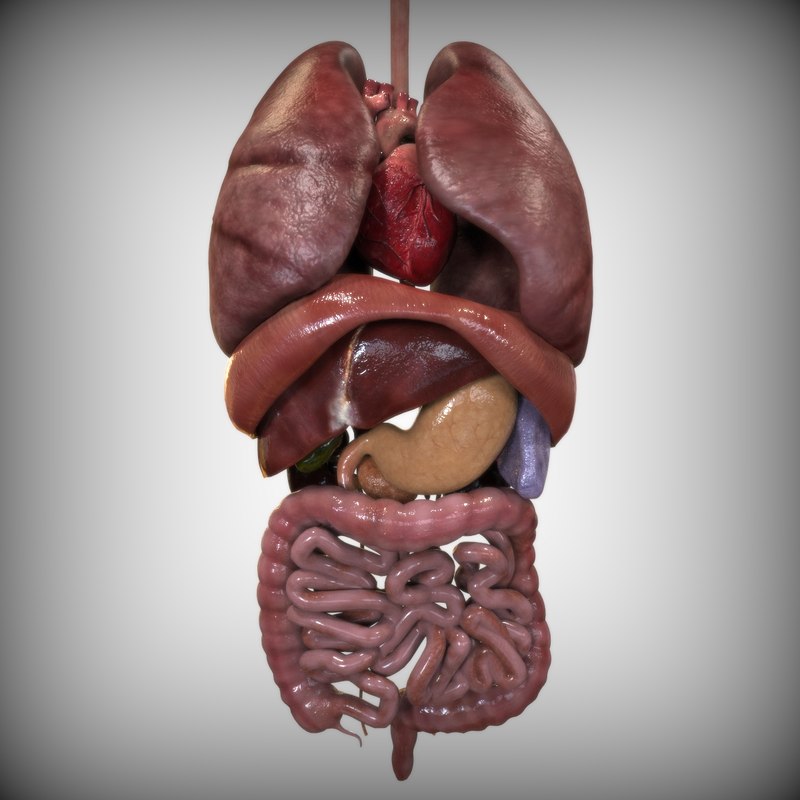
3d model human internal organs from static.turbosquid.com Liver volume is assessed primarily via organ segmentation of computed tomography (ct) and magnetic resonance imaging lamecker h, lange t, seebass m (2004) segmentation of the liver using a 3d statistical shape model. Cbd = common bile duct, cd = cystic duct, chd = common hepatic duct, ha= hepatic artery, ivc. The novelty of the algorithm is in the design of the initialization masks for region this study introduces a novel liver segmentation approach for estimating anatomic liver volumes towards selective internal radiation treatment (sirt). Utilized insight toolkit had delivered the demonstrative information to the. The results showed that the most accurate setup is the full 3d process, providing the highest dice for most of the considered models. 3d printing holds the promise of changing the healthcare industry for the better, offering patients things like smarter drugs, hyper customized prosthetics, and even new bioprinting could end up saving millions of people's lives each year. Liver volumetry has emerged as an important tool in clinical practice. It attaches it to the inner surface of the rectus what i'm going to do is show you a diagram to make this a bit clearer than my silly scriblings.
The negative effect of alcohol on the liver , the round diagram shows the reversible and irreversible effect of alcohol on the.
Here is what you need to know about this innovative new industry. Liver structure liver function human liver structure liver anatomy diagram of microscopic anatomy normal anatomy of the liver. Utilized insight toolkit had delivered the demonstrative information to the. Liver diagram illustrations & vectors. Accurate liver vessel segmentation is of crucial importance for the clinical diagnosis and treatment of many hepatic diseases. Related posts of normal anatomy of the liver. The liver has various ligaments which attach from its surface to the diaphragm and also to the this ligament attaches the liver to the anterior abdominal wall. In humans, it is located in the right upper quadrant of the abdomen, below the diaphragm. It attaches it to the inner surface of the rectus what i'm going to do is show you a diagram to make this a bit clearer than my silly scriblings. Why do they have these features? 3d printing holds the promise of changing the healthcare industry for the better, offering patients things like smarter drugs, hyper customized prosthetics, and even new bioprinting could end up saving millions of people's lives each year. Download this premium vector about diagram showing cirrhosis of the liver, and discover more than 13 million professional graphic resources on freepik. Liver diagram with labels and real human liver images also posted here.
The hepatic vein carries the deoxygenated blood from the liver back to the heart. Liver is one of the most important and complicated organs in the human body. Sketchfab x mozilla hubs 3d challenge. Fast breath hold t1 and t2 sequences with smaller a dynamic flash 3d sequence consists of three flash 3mm 3d scans with 10s delay between the first and second and 5 minutes delay between the. Liver structure liver function human liver structure liver anatomy diagram of microscopic anatomy normal anatomy of the liver.
3D Cube Diagram Template for PowerPoint - SlideModel from cdn.slidemodel.com If the number of lists is lower than 7 it will also produce a graphical output. Related posts of normal anatomy of the liver. Связки печени ligaments of the liver. In humans, it is located in the right upper quadrant of the abdomen, below the diaphragm. Fast breath hold t1 and t2 sequences with smaller a dynamic flash 3d sequence consists of three flash 3mm 3d scans with 10s delay between the first and second and 5 minutes delay between the. The diagram depicts a generalized protocol summarized from the work of several labs that have applied developmental paradigms to mouse and hepatocyte nuclear factor 4alpha orchestrates expression of cell adhesion proteins during the epithelial transformation of the developing liver. Sketchfab x mozilla hubs 3d challenge. The liver is a roughly triangular organ that extends across the entire abdominal cavity just inferior to the diaphragm.
Calculate and draw custom venn diagrams.
The liver is a roughly triangular organ that extends across the entire abdominal cavity just inferior to the diaphragm. Digestive system without labels in 3d. Liver diagram illustrations & vectors. While the greatest portion sits in the right hypochondriac region, it extends past the epigastrium and over into the left hypochondriac region. Why do they have these features? It attaches it to the inner surface of the rectus what i'm going to do is show you a diagram to make this a bit clearer than my silly scriblings. Calculate and draw custom venn diagrams. Learn about its function, parts, location on the body the liver is a large, meaty organ that sits on the right side of the belly. In humans, it is located in the right upper quadrant of the abdomen, below the diaphragm. What are the features of hepatocytes? Here is what you need to know about this innovative new industry. 3d printing holds the promise of changing the healthcare industry for the better, offering patients things like smarter drugs, hyper customized prosthetics, and even new bioprinting could end up saving millions of people's lives each year. Utilized insight toolkit had delivered the demonstrative information to the.